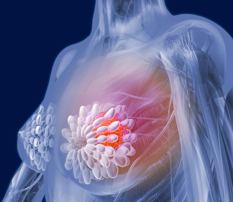
Breast cancer screening with mammography has been shown to improve prognosis and reduce mortality by detecting disease at an earlier stage. However, many cancers are missed on screening mammography.
Researchers now report that artificial intelligence (AI) may potentially improve reading breast cancer screening mammograms. Their findings, “Improving Breast Cancer Detection Accuracy of Mammography with the Concurrent Use of an Artificial Intelligence Tool,” was published recently in the journal Radiology: Artificial Intelligence.
“Mammography has been the frontline screening tool for breast cancer for decades with more than 200 million women being examined each year around the globe,” noted the researchers. “However, limitations in sensitivity and specificity persist even in the face of the most recent technologic improvements. Up to 30–40% of breast cancers can be missed during screening and on average, only 10% of women recalled from screening for diagnostic workup are ultimately found to have cancer…Our work described a multireader, multicase clinical investigation carried out to test the hypothesis that the use of a new AI system can improve the performance of radiologists in breast cancer detection when reading digital screening mammography.”
The researchers used MammoScreen, an AI tool by the company Therapixel that can be applied with mammography to aid in cancer detection. MammoScreen is designed to identify regions suspicious for breast cancer on two-dimensional digital mammograms, assess their likelihood of malignancy based on a complete set of four views, and generate a set of image positions with a related suspicion score.
The researchers assessed a dataset of 240 2D digital mammography images acquired between 2013 and 2016 that included different types of abnormalities. Half of the dataset was read without AI and the other half with the help of AI.
“The average area under the receiver operating characteristic curve (AUC) across readers was 0.769 (95% CI: 0.724, 0.814) without AI and 0.797 (95% CI: 0.754, 0.840) with AI. The average difference in AUC was 0.028 (95% CI: 0.002, 0.055, P = .035). Average sensitivity was increased by 0.033 when using AI support (P = .021). Reading time changed dependently to the AI-tool score. For low likelihood of malignancy (< 2.5%), the time was about the same in the first reading session and slightly decreased in the second reading session. For higher likelihood of malignancy, the reading time was on average increased with the use of AI,” the researchers wrote.
The reduced reading time could potentially increase overall radiologists’ efficiency, allowing them to focus their attention on the more suspicious examinations, the researchers said.

“The results show that MammoScreen may help to improve radiologists’ performance in breast cancer detection,” said Serena Pacilè, Ph.D., clinical research manager at Therapixel, where the software was developed.
According to Pacilè, the FDA cleared MammoScreen for use in the clinic, where it could help reduce the workload of radiologists. The researchers look forward to observing the behavior of the AI tool on a large screening-based population and its ability to detect breast cancer earlier.
Their research further reveals the the power of artificial intelligence and may one day lead to improved breast cancer detection and outcomes.


